Jiao-Kun Li1,2,
Xue-Duan Liu2,
Li Shen2,
Wei-Min Zeng2 ![]() ,
Guan-Zhou Qiu1
,
Guan-Zhou Qiu1
For correspondence:- Wei-Min Zeng Email: zengweimin1024@sina.com Tel:+8673188877472
Received: 4 August 2015 Accepted: 31 March 2016 Published: 27 May 2016
Citation: Li J, Liu X, Shen L, Zeng W, Qiu G. Natural plant polyphenols for alleviating oxidative damage in man: Current status and future perspectives. Trop J Pharm Res 2016; 15(5):1089-1098 doi: 10.4314/tjpr.v15i5.27
© 2016 The authors.
This is an Open Access article that uses a funding model which does not charge readers or their institutions for access and distributed under the terms of the Creative Commons Attribution License (http://creativecommons.org/licenses/by/4.0) and the Budapest Open Access Initiative (http://www.budapestopenaccessinitiative.org/read), which permit unrestricted use, distribution, and reproduction in any medium, provided the original work is properly credited..
The balance between oxidation and reduction is important for maintaining a healthy biological system. Oxidative stress results from an imbalance between excessive formation of reactive oxygen species (ROS) and/or reactive nitrogen species (RNS) and limited endogenous defense systems, and this imbalance can adversely alter lipids, proteins and DNA, causing a number of human diseases. Thus, exogenous antioxidants that can neutralize the effect of free radicals are needed to diminish the cumulative effects of oxidative damage over human life span. Current research reveals that phenolic compounds in plants possess high antioxidant activity and free radical scavenging capacity and can prevent the body from oxidative damage over human life span. This review focuses on the present understanding of free radicals and antioxidants and their importance in human health and disease. Information about the chemical features of free radicals as well as their deleterious effects on cell structures is reviewed. The chemical structure and anti-oxidative mechanisms of essential polyphenols and their potential health benefits are presented. In addition, the limitation of natural antioxidants and a perspective on likely future trends in this field are also discussed.
Introduction
Oxidation is a chemical reaction involving transfer of an electron from electron - rich to electron - deficient entity. In human body, the oxygen molecule (O2) is an element indispensable for life and used biologically to oxidize (burn) carbon-and hydrogen-rich molecules to obtain chemical energy and heat [1]. However, oxygen is a dangerous friend. The by-products of its metabolism called free radicals, usually including reactive oxygen species (ROS) and reactive nitrogen species (RNS) are unstable, violently reactive and potentially destructive [2]. Although ROS and RNS exert beneficial effects on cellular responses and immune function at low/moderate levels, they also can damage all cell structures at high concentrations (termed as oxidative stress), leading to a number of diseases such as cancer, aging, cardiovascular and neurodegenerative diseases [3,4].
Antioxidants are substances capable of preventing or slowing the oxidation of other molecules. They exert their protective action either by suppressing the formation of free radicals or by scavenging free radicals [5]. However, oxidative stress occurs in the body when there is a serious imbalance between the generation of free radicals and the antioxidant defence systems [6]. Despite the fact that humans naturally produce antioxidants, the process is not effective under some physiopathological situations (cigarette smoke, air pollutants, UV radiation, inflammation) [7,8]. Thus, increasing the antioxidant intake can prevent diseases and lower the health problems caused by free radicals. Plants, especially the medicinal plants are important sources of antioxidant substances, which can inhibit free radical formation and/or interrupt propagation of autoxidation. In recent years, plant polyphenols have gained a lot of importance because of their potential use as prophylactic and therapeutic agents in many diseases related to oxidative stress and free radical-induced damage.
BASICS OF FREE RADICAL RESEARCH
Free radicals
The term ‘free radicals’ is defined as the reactive molecular species that contain unpaired electrons in their outermost orbital [2,3]. Free radicals can be formed from molecules by the homolytic fission of a chemical bond and via redox reactions, which is a far more common process in biological systems [2]. In popular scientific/biomedical literature, ‘free radical’ is used in a broad sense and also includes related reactive species such as ‘excited states’ that lead to free radical generation or those species that result from free radical reactions. Usually, free radicals are very short - lived and derived from two elements: oxygen and nitrogen, thus creating highly reactive molecules like reactive oxygen species (ROS) and reactive nitrogen species (RNS). ROS include superoxide anion radicals (O2‒•), reactive hydroxyl radicals (OH•), hydroperoxyl radical (HO2•) and other species like hydrogen peroxide (H2O2), hypochlorous acid (HOCl) and singlet oxygen (1O2) [9]. The nitrogen-derived free radicals are nitric oxide (NO•), nitrogen dioxide (NO2), peroxy-nitrite anion (ONOO–) [10].
Oxidative damages induced by free radicals
Although structurally different, the presence of unpaired electron results in free radicals sharing common properties, oxidizing other molecules to gain electrons and stabilizing themselves. Free radicals can attack important macromolecules in the human body, including lipids, proteins and DNA [11], and have been implicated as the cause of cancer, aging and neurodegenerative diseases such as Alzheimer’s disease, Parkinson disease and cardiovascular diseases such as arteriosclerosis. They are also the primary cause of cell death and tissue damage resulting from heart attack and stroke [12]. Lipids are more susceptible to free radical damage among all the major classes of biomolecules. When they react with membrane lipids, free radicals cause lipid peroxidation (LP). The consequences of lipid peroxidation are cross linking of membrane proteins, change in membrane fluidity and formation of adducts with protein and DNA which may be detrimental to the functioning of the cell [13]. In addition, a large number of toxic products are also formed due to lipid peroxidation, such as malondialdehyde (MDA), 4-hydroxynonenal (4-HNE) and various 2-alkenals.
Proteins can be oxidatively modified in three distinct ways: oxidative modification of a specific amino acid, free radical-mediated peptide cleavage, and formation of protein cross-linkage due to reaction with lipid peroxidation products [14]. The side chains of all amino acid residues of proteins, in particular tryptophan, cysteine and methionine residues are susceptible to oxidation [15]. Free radical mediated protein modification increases susceptibility to enzyme proteolysis. In addition, oxidative damage to cellular proteins can affect the signal transduction pathways, enzyme activity and heat stability which leads to aging [16].
Although DNA is a stable, well-protected molecule, ROS/RNS still can interact with it and cause several types of damage such as production of base-free sites, deletions, modification of all bases, frame shifts, double strand DNA breaks, DNA-protein cross-links and chromosomal arrangements [17]. Because oxidative damage to DNA can affect the cell cycle and lead to mutations, DNA alteration has been suggested to be responsible in part in carcinogenesis [18,19].
Source of free radicals in human body
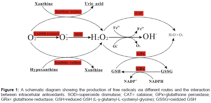
Free radicals and other ROS are derived either from endogenous metabolic process in the human body or from external sources, such as exposure to X-rays, ozone, cigarette smoking, air pollutants, and industrial chemicals. In the cells, formation of ROS and RNS can occur continuously as a consequence of both enzymatic and non-enzymatic reactions. Enzymatic reactions generating free radicals include those involved in the respiratory chain, in phagocytosis, in the prostaglandin synthesis, and in the cytochrome P450 system [20]. Mitochondria have long been recognized as the major site for ROS production and both complexes I and III have been established to be the specific sites for mitochondrial ROS generation [21,22]. Besides mitochondria, many enzymes are also capable of producing ROS, such as, NADPH oxidase, xanthine oxidase, D-amino acid oxidase and dihydrolipoamide dehydrogenase [23]. For example, the superoxide anion radicals (O2‒•) is generated via cellular oxidase systems, such as NADPH oxidase, xanthine oxidase, peroxidases. Hydrogen peroxide (H2O2) is produced by the action of several oxidase enzymes, including amino acid oxidase and xanthine oxidase. In particular, xanthine oxidase catalyzes the reaction of hypoxanthine to xanthine and xanthine to uric acid. In both steps, molecular oxygen is reduced, forming the superoxide anion in the first step and hydrogen peroxide in the second [20-22]. Hydroxyl radical (OH•), the most reactive free radical is formed by the reaction of O2‒• with H2O2 in the presence of Fe2+ or Cu+ (the Fenton reaction) [24]. The various pathways involved in the generation of some of the reactive oxygen species are given in . In addition, free radicals can also be formed in non-enzymatic reactions of oxygen with organic compounds as well as those initiated by ionizing radiations.
Oxidative stress and antioxidant protection mechanisms
In general, free radicals as necessary intermediates are produced in a variety of normal biochemical reactions and a homeostatic balance exists between free radical generation and quenching under normal physiological conditions [25,26]. Oxidative stress occurs when this balance is disrupted by excessive production of reactive oxygen species. Antioxidants are central to the redox balance in the human body. The term ‘antioxidant’ refers to any molecule stable enough to donate an electron to a rampaging free radical and neutralize it, thus reducing its capacity to damage a target molecule [2,3]. Antioxidants may exert their effects by different mechanisms, such as suppressing the production of active species by reducing hydroperoxides and H2O2 and also by sequestering metal ions, termination of chain reaction by scavenging active free radicals, repairing and/or clearing damage of cell. Similarly, some antioxidants also induce the biosynthesis of other antioxidants or defence enzymes [27].
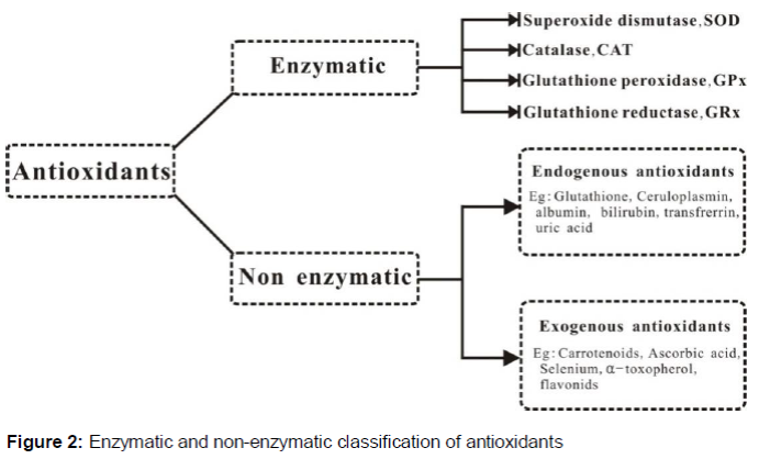
Humans have several mechanisms to counteract oxidative stress, either by producing antioxidants from endogenous antioxidant systems or externally supplied through exogenous antioxidants (). The endogenous antioxidant systems, including enzymatic and non-enzymatic antioxidants, play a crucial role in maintaining optimal cellular functions. The major antioxidant enzymes directly involved in the neutralization of ROS and RNS are superoxide dismutase (SOD), catalase (CAT), glutathione peroxidase (GPx), glutathione reductase (GRx) [3,28]. SOD, the first line of defense against free radicals, catalyzes the dismutation of O2‒• to O2 and to the less-reactive species H2O2 by reduction. In humans there are three forms of SOD: cytosolic Cu, Zn-SOD, mitochondrial Mn-SOD, and extracellular SOD (EC-SOD) [29]. The H2O2 is transformed into water and oxygen by CAT or GPx. The selenoprotein GPx enzyme removes H2O2 by using it to oxidize reduced glutathione (GSH) into oxidized glutathione (GSSG). Glutathione reductase, a flavoprotein enzyme, regenerates GSH from GSSG, with NADPH as a source of reducing power ().
The non-enzymatic antioxidants are also divided into endogenous (metabolic) and exogenous (nutrient) antioxidants. Endogenous antioxidants are produced by metabolism in the body, such as lipoid acid, glutathione, L-ariginine, coenzyme Q10, melatonin, uric acid, bilirubin, metal-chelating proteins, transferrin [30]. Exogenous antioxidants are compounds that cannot be produced in the body, such as vitamin E, vitamin C, carotenoids, trace metals (selenium, manganese, zinc), flavonoids, omega-3 and omega-6 fatty acids. These exogenous antioxidants must be provided by foods or supplements via diet.
PLANTS AS SOURCES OF NATURAL ANTIOXIDANTS
Despite the fact that humans are equipped with an impressive repertoire of antioxidant enzymes as well as small antioxidant molecules, these agents may not be sufficient enough to normalize the redox status during oxidative stress [31]. Antioxidant supplementation/treatment has been adopted for maintaining optimal cellular functions. There are a number of synthetic phenolic antioxidants that have been widely used as food antioxidants, such as butylated hydroxyanisole (BHA), butylated hydroxytoluene (BHT) and ter-butylhydroquinone (TBHQ) [32]. However, some physical properties of synthetic antioxidants, such as their high volatility and instability at elevated temperatures, carcinogenic nature and consumers’ preference for natural ingredients have reinforced the efforts for the development of alternative antioxidants from natural origins.
Plants, especially medicinal herbs, have been used for the prevention and/or treatment of several diseases since very old times [33]. Plant extracts, such as flavonoids and phenolics, have raised public interest in their potential to act as antioxidants. Natural antioxidants can strengthen the endogenous antioxidant defense from ROS ravage and restore the optimal balance by neutralizing the reactive species [34]. In traditional Chinese medicine (TCM), a similar concept of balance between anti-oxidation and oxidation called yin-yang has existed for more than 2000 years [35]. ‘Yang-invigorating’ action usually associates with immune-enhancement and energy generation enhancement, i.e., through the enhancement of the mitochondrial oxidative process, while ‘yin-nourishing’ action suppress the symptom of heat-fire or ‘yang’ i.e., preventing the over oxidation process [36]. Maintaining yin and yang in harmony is akin to attaining the homeostatic state. An imbalance between ROS and the inherent antioxidant capacity of the body, has directed the use of dietary and/or medicinal supplements particularly during the disease attack [36]. The epidemiological studies have demonstrated an inverse association between ingestion of these natural antioxidants and mortality from age-related diseases, such as coronary heart diseases and cancer [37]. Based on a recent large-scale research [38], large number of medicinal plants has been identified as having potential antioxidant activities [39-42]. The raw extracts or isolated pure compounds from medicinal plants are more effective antioxidants in vitro than BTH or vitamin E [43,44]. Moreover, the medicinal plants also exhibit far stronger antioxidant activity and contain significantly higher levels of phenolic compounds than common vegetables and fruits [38]. Therefore, the medicinal plants are promising sources of natural antioxidants [45].
Structures and classes of polyphenols
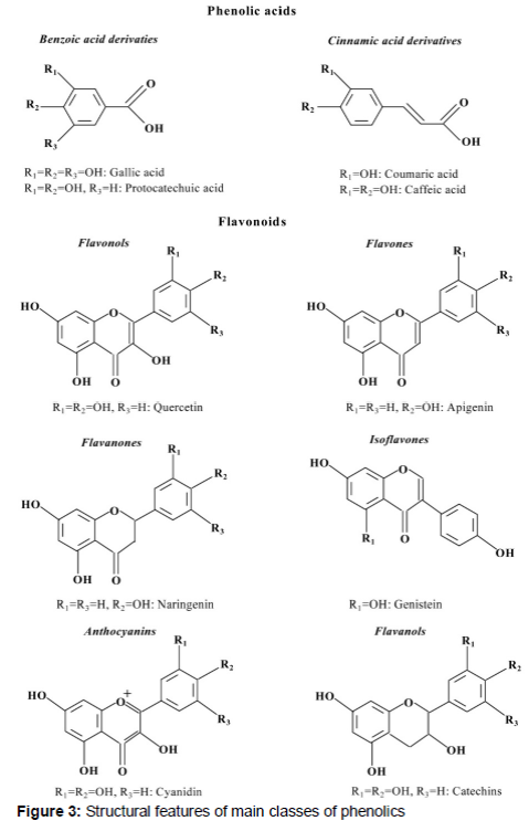
Polyphenols are secondary metabolites of plants and generally involved in defense against ultraviolet radiation or aggression by pathogens [46]. They comprise a wide variety of molecules that have polyphenol structures, i.e. several hydroxyl groups on aromatic rings, but also molecules with one phenol rings, such as phenolic acid and phenolic alcohols. According to the number of phenol rings that they contain and to the structural elements that bind these rings to one another, polyphenols are classified into different groups, including phenolic acids, flavonoids, stilbenes and lignans [47]. Flavonoids and phenolic acids have been considered as the major contributors to the antioxidant activity in medicinal plants [47].
Flavonoids comprise the most abundant group of plant polyphenols [48]. Their common structural feature is the diphenylpropane moiety, which consists of two aromatic rings linked through three carbon atoms that together usually form an oxygenated heterocycle (). Based on the variation in the type of heterocycle involved, flavonoids are divided into six classes: flavones, flavanones, flavonols, isoflavones, anthocyanidins and flavanols (or catechins) (). Flavonols are the most ubiquitous flavonoids in foods with quercetin and kaempferol as the more representative compounds. Phenolic acids can be divided in two classes: derivatives of benzoic acid and derivatives of cinnamic acid (). The hydroxybenzoic acids, such as gallic acid and protocatechuic acid, are found in very few edible plants, except for certain red fruits, black radish, and onions [49]. The hydroxycinnamic acids are more common than are the hydroxybenzoic acids and consist chiefly of p-coumaric, caffeic, ferulic, and sinapic acids. Gallic acid, the precursor of many tannins, is one of the most studied and promising compounds in the hydroxybenzoic group, while cinamic acid is the precursor of all the hydroxycinnamic acids [50].
CLINICAL EFFECTS OF POLYPHENOLS IN OXIDATIVE STRESS-RELATED DISEASES
It is well established that consumption of polyphenol-rich foods may increase plasma antioxidant capacity [51,52]. This increase in the anti-oxidative capacity of plasma may be explained either by the presence of reducing polyphenols and their metabolites in plasma, by their effects upon concentrations of other reducing agents (endogenous antioxidants), or by their effect on the absorption of pro-oxidative food components, such as iron [47]. Epidemiological studies have repeatedly shown that polyphenols, as antioxidants may protect cell constituents against oxidative damage and therefore limit the risk of various degenerative diseases associated with oxidative stress [53].
Polyphenols and aging
Aging can generally be defined as a progressive decline in the efficiency of biochemical and physiological processes after the reproduction phase of life. It is a natural accumulation process of diverse detrimental changes in cells and tissues, leading to the disabilities and diseases that limit normal body functions [54]. Among the theories proposed for the explaining the mechanism of aging [55-57], free radical/oxidative stress theory is one of the most accepted [58]. Although a certain amount of oxidative damage takes places under normal conditions, the rate of this damage significantly increases during the aging process as the efficiency of anti-oxidative and repair mechanisms decrease [59].
Anthocyanins are particularly abundant in brightly colored fruits such as berry fruits and concord grapes and grape seeds. They have been shown to have potent antioxidant/anti-inflammatory activities, as well as to inhibit lipid peroxidation and the activity of cyclo-oxygenase (COX)-1 and -2 [60]. Fruit and vegetable extracts that have high levels of flavonoids also display high total antioxidant activity. It is reported that the dietary supplementation with spinach, strawberry or blueberry extracts in a control diet were effective in reversing aging-related deficits in brain and behavioral function in aged rats [61]. Tea catechins have been shown to possess strong anti-aging activity and consuming green tea rich in these catechins may delay the onset of aging [62]. Resveratrol is a very recent entry as an anti-aging agent for preventing oxidative stress-induced aging in endothelia cells by preventing ROS-induced damage via increased expression of endothelia silencing information regulator (SIRT1). SIRT1 belongs to the nicotinamide adenine dinucleotide (NAD+) dependent histone deacetylase family regulates gene silencing and is believed to mediate the beneficial effects on health and longevity in normal cells by calorie restriction [63].
Polyphenols and cardiovascular disease
Cardiovascular disease (CVD) is one of the leading causes of death in many developed nations [64]. CVD is a chronic, multi-factorial disease in which a range of genetic and environmental factors plays a role in its initiation, progression and development [65,66]. Arteriosclerosis (hardening of the arteries), one of the classical examples of an ROS-mediated CVD, is the result of oxidative modification of low density lipoprotein (LDL) in the arterial walls [67]. Many human studies have showed an association of moderate intake of alcoholic drinks containing polyphenols with a reduced risk of coronary heart diseases [68-70]. The mechanisms by which polyphenols may be protective against CVD are inhibiting LDL oxidation inhibiting platelet aggregation and reducing inflammatory response [71,72].
Resveratrol is the active compound in red wine which is attributed for “French Paradox”, the low incidence of CVD despite the intake of high-fat diet and smoking among the French [73]. Studies have shown that resveratrol potentially inhibits the oxidation of the LDL particles via chelating copper or by direct scavenging of the free radicals [74,75]. Meanwhile, resveratrol can prevent platelet aggregation via preferential inhibition of COX-1 activity, which synthesizes thromboxane A2, an inducer of the platelet aggregation and vasoconstrictor [76]. In addition, quercetin has been shown to be inversely associated with mortality from coronary heart disease by inhibiting the expression of metalloproteinase1 (MMP1), and the disruption of atherosclerotic plaques [72].
Polyphenols and cancer
Carcinogenesis is a multistage process and can be divided into three stages: initiation, promotion and progression. ROS can act in all these stages of carcinogenesis [77]. The anticancer effects of polyphenols have been observed at various sites, such as mouth, stomach, duodenum, colon, liver, lung, mammary gland and skin. Many polyphenols, such as quercetin, catechins, isoflavones, ligans, flavanones, ellagic acid, resveratrol have been tested; all of them showed protective effects in some models although their mechanism of action were found to be different [78].
Several mechanisms of action have been identified for anticancer effects of polyphenols. These include removal of carcinogenic agents [79], modulation of cancer cell signaling [80] cell cycle progression [81], promotion of apoptosis [82] and modulation of enzymatic activities [83]. For example, polyphenols influence the metabolism of pro-carcinogens by modulating the expression of cytochrome P450 enzymes involved in their activation to carcinogens. Furthermore, they may modulate the activity of signaling pathways, such as MAPK kinase and PI3 kinase, which are involved in cancer proliferation [84].
Polyphenols and neurodegeneration
Neurodegenerative disorders such as Parkinson’s and Alzheimer’s diseases represent an increasing problem in our aging societies [85]. Mitochondrial dysfunction and oxidative damage have been identified as the risk factors for neurodegenerative diseases [85]. Because polyphenols are highly antioxidative in nature, their consumption may provide protection in neurological diseases. For example, polyphenol-rich Ginkgo biloba extracts have been shown to be neuroprotective by protecting hippocampal neurons from nitric oxide- and beta-amyloid-induced neurotoxicity [86]. Anthocyanins and isoflavones can reduce the neurodegeneration associated with accumulation advanced glycation endproducts (AGEs) during normal and abnormal brain aging [87]. In addition, phenolic compounds such as caffeic acid and tyrosol are capable of inducing neuroprotective effects to a similar extent to that seen with flavonoids [88].
Conclusion
There is no doubt that the correct balance between oxidation and reduction is critical in maintaining a healthy biologic system. In this review, we have summarized the chemical features of free radicals as well as their deleterious effects on cell structures. In addition, the potential roles of the natural antioxidants from medicine plants in preventing and repairing damages caused by oxidative stress are presented. For a proper evaluation of their potential health effects, we need more data on the concentrations and metabolic forms that tissues and cells are exposed to after ingestion of plant polyphenols via the diet. In addition, new genomic techniques will give tremendous opportunities to explore this field. The high-through genomics tools will then be able to increase our understanding on how flavonoids affect metabolic pathways and, as a consequence, affect human health.
Declarations
Acknowledgement
References
Archives


News Updates